
Anticorps > 20231 - Purified freeze-dried antibody to chicken type II collagen
20231 - Purified freeze-dried antibody to chicken type II collagen
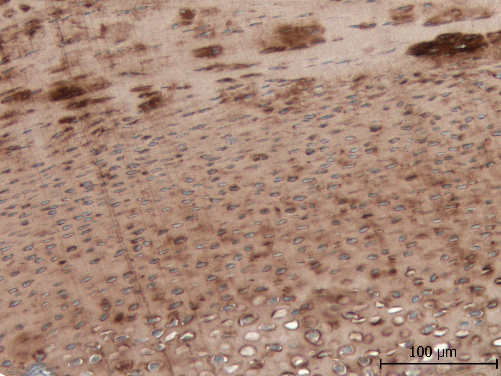
Chicken cartilage paraffin section, IHC
species
Chicken
immunogen
Type II collagen
Type II collagen is a fibrillar collagen composed of three identical α1(II) chains. The type II collagen is found in cartilage and the vitreous humor of the eye. Mutations in this gene are associated with achondrogenesis, chondrodysplasia, early onset familial osteoarthritis, SED congenita, Langer-Saldino achondrogenesis, Kniest dysplasia, Stickler syndrome type I, and spondyloepimetaphyseal dysplasia Strudwick type. [according to RefSeq]
Immunogen : Native type II collagen extracted from chicken cartilage.
Host : Rabbit.
Polyclonal antibody purified by chromatography.
For research only.
Purified, freeze-dried antibody in 0.1 mL vials. Reconstitute with 0.1 mL distilled water and store aliquots at -20°C.
Unreconstituted
24 months at -20°C.
Reconstituted
Before use aliquot and store at -20°C (6 months). Avoid repeated freeze/thaw cycles.
Applications
IF, IHC, ELISA
Working dilutions
ELISA ≥ 1/2000 (OD = 0.5)
IF ≥ 1/40
IHC ≥ 1/500
Optimal dilutions should be determined by the end user.
- #318j
-
IgG
4,4 mg/mL
pH
7,8
ELISA
ELISA not tested with our current protocol.
IF
With fluorescein anti-rabbit IgG conjugate, optimal working dilution at 1/40 on frozen chicken cartilage tissues.
IHC
Immunoperoxidase technique with the Envision / Dako kit, 0.5% hyaluronidase pretreatment, optimal working dilution at 1/500 on fixed paraffin-embedded chicken cartilage.
-
Presentation
Type II collagen is a fibrillar collagen composed of three identical α1(II) chains. The type II collagen is found in cartilage and the vitreous humor of the eye. Mutations in this gene are associated with achondrogenesis, chondrodysplasia, early onset familial osteoarthritis, SED congenita, Langer-Saldino achondrogenesis, Kniest dysplasia, Stickler syndrome type I, and spondyloepimetaphyseal dysplasia Strudwick type. [according to RefSeq]
Description
Immunogen : Native type II collagen extracted from chicken cartilage.
Host : Rabbit.
Polyclonal antibody purified by chromatography.
For research only.
Format
Purified, freeze-dried antibody in 0.1 mL vials. Reconstitute with 0.1 mL distilled water and store aliquots at -20°C.
Stability
Unreconstituted
24 months at -20°C.
Reconstituted
Before use aliquot and store at -20°C (6 months). Avoid repeated freeze/thaw cycles.
Use
Applications
IF, IHC, ELISA
Working dilutions
ELISA ≥ 1/2000 (OD = 0.5)
IF ≥ 1/40
IHC ≥ 1/500
Optimal dilutions should be determined by the end user.
Lots
- #318j
-
IgG :
4,4
pH :
7,8
ELISA
ELISA not tested with our current protocol.
IF
With fluorescein anti-rabbit IgG conjugate, optimal working dilution at 1/40 on frozen chicken cartilage tissues.
IHC
Immunoperoxidase technique with the Envision / Dako kit, 0.5% hyaluronidase pretreatment, optimal working dilution at 1/500 on fixed paraffin-embedded chicken cartilage.
-
References
- «Immunohistochemical study of collagens of the extracellular matrix in cartilage of Sepia officinalis. Eur. J. Histochem. 1999, 43, 211-225.»
Bairati A., Comazzi M., Gioria M., Hartmann D.J., Leone F., Rigo C. - «Processing of Type XI Collagen. J. Biol. Chem., 1996, 271, 23743-23748.»
Rousseau J.C., Farjanel J., Boutillon M.M., Hartmann D.J., van der Rest M., Moradi-Ameli M. - «Bone organotypic culture method: a model for cytocompatibility testing of biomaterials. Cells and Materials 1994, 4, 113-123.»
Anselme K., Lanel B., Gentil C., Hardouin P., Marie P.J., Sigot-Luizard M.F. - «Proteglycan and collagen synthesis are correlated with actin organization in dedifferentiating chondrocytes. Eur. J. Cell. Biol. 1991, 56, 364-373.»
Mallein-Gerin F., Garrone R., Van der Rest M. - «Proteoglycan core protein and type II collagen gene expressions are not correlated with cell shape changes during low density chondrocyte cultures. Differentiation 1990, 43, 204-211.»
Mallein-Gerin F., Ruggiero F., Garrone R. - «In vitro morphogenesis of chick embryo hypertrophic cartilage. J. Cell. Biol., 1987, 105, 999-1006.»
Tacchetti C., Quarto R., Nitsch L., Hartmann D.J., Cancedda R.