Services > RNAscope®
RNAscope®
Next-Generation In Situ Hybridization: RNAscope® Technology Take your tissue characterization to the next level.
Novotec is expanding its in situ molecular biology services by integrating RNAscope® technology. This cutting-edge, highly sensitive, and specific in situ hybridization (ISH) method overcomes the limitations of traditional ISH techniques. From gene to protein, Novotec provides absolute-precision cellular mapping to help you fully understand complex biological mechanisms.
Why choose RNAscope® technology ?
RNAscope® relies on patented "Z" probe design and signal amplification technology, offering major advantages over conventional ISH:
-
Exceptional Sensitivity: Enables detection of single RNA molecules (single-molecule RNA) that are highly degraded or expressed at extremely low levels.
-
Maximum Specificity: The amplification system is only triggered when two independent target probes bind side by side, effectively eliminating background noise.
-
Preserved Morphology: Allows visualization of signals as distinct dots on formalin-fixed paraffin-embedded (FFPE) or frozen tissue sections.
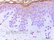

Novotec Innovation: RNAscope® + IHC/IF co-staining
For absolute spatial understanding, we combine the detection of target RNA transcripts and proteins on the exact same tissue section—whether for the same gene product or distinct targets:
-
Dual Chromogenic ISH/IHC: Our validated protocol pairs RNAscope® with classic immunohistochemistry. This allows you to correlate gene expression with protein translation while precisely identifying the specific cell type expressing your RNA of interest using phenotypic markers (surface proteins, intermediate filaments, etc.).
-
Immunofluorescence (IF) Developments: True to its innovative spirit, our R&D team is currently developing fluorescent co-staining to pave the way for even finer multiplex analysis, facilitating the study of complex interactions between different genes and proteins within the tissue microenvironment.
Application & Research fields
-
Xenograft Monitoring & Cell Persistence: Precise mapping and differentiation of human cell persistence within host tissues of different species (animal models).
-
3D Model Biology (Spheroids & Organoids): Phenotypic monitoring, characterization, and validation of cell identity retention within 3D structures.
-
Cosmetic Screening & Treatment Evaluation: Precise measurement of the impact of formulations or physical stressors (e.g., UV irradiation) on RNA modulation, RNA synthesis, and protein expression in reconstructed skin models.
-
Dermatology & Tissue Pathologies: Fine-grained analysis of inflammatory and pathophysiological mechanisms (e.g., dermatitis, psoriasis) at the cellular level.


References
- «In situ Hybridization to evaluate the wound healing process of a bilayered living cell-based construct (Apligraf®) in a nude mice model - 2010 june, TERMIS-EU, Galway, Ireland.»
Rousseau C. and Pagnon-Minot A., Guerret S., Hartmann D.J, Ronfard V. - «Short term integration on human bilayered living skin substitute (Apligraf®) grafted onto nude mice - 19th Annual Meeting of the Wound Healing Society with SAWC (Symposium on Advanced Wound Care) 2009 April 26-29, Gaylord Texan Resort and Convention Center, Dallas, Texas.»
Rousseau C., Thillou F., Pagnon-Minot A., Guerret S., Hartmann D.J, Ronfard V. - «Characterization of spatial and temporal expression of pattern of Col15a1b during zebrafish development. Gene Expression Patterns 2011, 11, 129-134.»
Bretaud S., Pagnon-Minot A.,Guillon E., Ruggiero F., Le Guellec D. - «Collagen XV, a novel factor in zebrafish notochord differentiation and muscle development. Dev Biol 2008, 316, 21-35.»
Pagnon-Minot A., Malbouyres M., Haftek-Terreau Z., Kim H.R., Sasaki T., Thisse C., Thisse B., Ingham P.W., Ruggiero F., Le Guellec D.
